Treating Mortons Neuroma

Overview

The nerves located on the bottom of the foot between the metatarsal heads (the bone of the toe closest to the foot) supply feeling, or sensation, to the toes. One of these nerves may become irritated by the ligament located above it, causing it to become thickened and painful. Generally this problem occurs most often between the third and fourth toes, but it can occur between the second and third toes as well. It is uncommon for a neuroma to be located between the first and second or fourth and fifth metatarsals. Rarely is there more than one neuroma in a foot.

Causes

Morton’s neuroma is an inflammation caused by a buildup of fibrous tissue on the outer coating of nerves. This fibrous buildup is a reaction to the irritation resulting from nearby bones and ligaments rubbing against the nerves. Irritation can be caused by wearing shoes that are too tight, wearing shoes that place the foot in an awkward position, such as high heels, a foot that is mechanically unstable, repetitive trauma to the foot such as from sports activities like tennis, basketball, and running. Trauma to the foot caused by an injury such as a sprain or fracture. It is unusual for more than one Morton’s neuroma to occur on one foot at the same time. It is rare for Morton’s neuroma to occur on both feet at the same time.

Symptoms

Pain is usually increased by forefoot weight bearing activities (such as running), with narrow-fitting footwear, or with high heeled shoes. It is usually painful to firmly touch the affected region and, in chronic cases, pain and sometimes an audible click, may be heard when squeezing the foot and toes together with the hand. Often a localized area of swelling may be evident at the site of injury.

Diagnosis

You should visit a doctor or podiatrist (foot doctor) if you have pain or tingling that does not stop. Your health care provider will examine your feet and will apply pressure on the spaces between the bones of the toes to determine the location of the foot pain. The doctor may order X-rays to rule out other conditions associated with foot pain, such as a stress fracture or arthritis. X-rays alone will not show whether or not a neuroma is present, so an ultrasound scan or magnetic resonance imaging (MRI) test may be done to confirm the diagnosis. A diagnostic procedure called an electromyography is sometimes used to rule out nerve conditions that may cause symptoms like those of associated with Morton?s neuroma.

Non Surgical Treatment

Nonsurgical treatment is tried first. Your doctor may recommend any of the following. Padding and taping the toe area, shoe inserts, changes to footwear, for example wearing shoes with wider toe boxes or flat heels, Anti-inflammatory medicines taken by mouth or injected into the toe area, nerve blocking medicines injected into the toe area, other painkillers, physical therapy. Anti-inflammatories and painkillers are not recommended for long-term treatment. In some cases, surgery is needed to remove the thickened tissue and inflammed nerve. This helps relieve pain and improve foot function. Numbness after surgery is permanent.intermetatarsal neuroma

Surgical Treatment

Operative treatment of Morton?s neuroma should be entertained only after failure of non-operative management. Standard operative treatment involves identifying the nerve and cutting (resecting) it proximal to the point where it is irritated/injured. This is usually done through an incision on the top (dorsal) aspect of the foot, although an incision on the sole (plantar) aspect of the foot can be used. Some physicians will attempt to treat Morton?s neuroma by releasing the intermetatarsal ligament, and freeing the nerve of local scar tissue. This may also be beneficial.

Prevention

Although the exact causes of neuromas are not completely known, the following preventive steps may help. Make sure your exercise shoes have enough room in the front part of the shoe and that your toes are not excessively compressed. Wear shoes with adequate padding in the ball of the foot. Avoid prolonged time in shoes with a narrow toe box or excessive heel height (greater than two inches).

What Are The Main Causes Of Heel Spur

Heel Spur

Overview

One of the conditions of the heel that can cause a lot of inconvenience is the development of heel spurs. A heel spur is the growth of calcium deposit on the heel bone. This deposit can become a bony protrusion and can cause a great deal of discomfort and pain when standing or walking.

Causes

A major cause of heel spur pain comes from the development of new fibrous tissue around the bony spur, which acts as a cushion over the area of stress. As this tissue grows, a callus forms and takes up even more space than the heel spur, leading to less space for the thick surrounding network of tendons, nerves, ligaments and supporting tissue. These important structures in the foot have limited space because of calcium or tissue buildup, which leads to swelling and redness of the foot, and a deep throbbing pain worsened with exercise.

Heel Spur

Symptoms

Heel spurs result in a jabbing or aching sensation on or under the heel bone. The pain is often worst when you first arise in the morning and get to your feet. You may also experience pain when standing up after prolonged periods of sitting, such as work sessions at a desk or car rides. The discomfort may lessen after you spend several minutes walking, only to return later. Heel spurs can cause intermittent or chronic pain.

Diagnosis

Heel spurs and plantar fasciitis are diagnosed based on the history of pain and tenderness localized to these areas. They are specifically identified when there is point tenderness at the bottom of the heel, which makes it difficult to walk barefoot on tile or wood floors. X-ray examination of the foot is used to identify the bony prominence (spur) of the heel bone (calcaneus).

Non Surgical Treatment

Get some rest. You need to stay off of your aching foot as much as possible for at least a week. Think about possible causes of the problem while you’re resting and figure out how you can make some changes. Some actions that can contribute to heel spurs include running too often or running on hard surfaces such as concrete, tight calf muscles, shoes with poor shock absorption. Ease back into your activities. In many cases, you’ll be in too much pain to go ahead with a strenuous exercise routine that puts pressure or impact on your heel. Listen to your body and switch to different activities such as swimming or riding a bike until your heel spurs improve.

Surgical Treatment

Surgery involves releasing a part of the plantar fascia from its insertion in the heel bone, as well as removing the spur. Many times during the procedure, pinched nerves (neuromas), adding to the pain, are found and removed. Often, an inflamed sac of fluid call an accessory or adventitious bursa is found under the heel spur, and it is removed as well. Postoperative recovery is usually a slipper cast and minimal weight bearing for a period of 3-4 weeks. On some occasions, a removable short-leg walking boot is used or a below knee cast applied.

Prevention

To prevent this condition, wearing shoes with proper arches and support is very important. Proper stretching is always a necessity, especially when there is an increase in activities or a change in running technique. It is not recommended to attempt working through the pain, as this can change a mild case of heel spurs and plantar fascitis into a long lasting and painful episode of this condition.

What Are The Primary Causes Of Posterior Calcaneal Spur

Calcaneal Spur

Overview

If you’re feeling pain on the bottom of your foot near your heel, pain after exercise or activity, or pain first thing in the morning or after a long period of sitting, then you may have a heel spur. Heel spurs don’t have a magic cure, but you can take steps to ease the pain and to eventually get rid of them.

Causes

At the onset of this condition, pain and swelling become present, with discomfort particularly noted as pushing off with the toes occurs during walking. This movement of the foot stretches the fascia that is already irritated and inflamed. If this condition is allowed to continue, pain is noticed around the heel region because of the newly formed bone, in response to the stress. This results in the development of the heel spur. It is common among athletes and others who run and jump a significant amount.

Posterior Calcaneal Spur

Symptoms

Heel spur is characterised by a sharp pain under the heel when getting out of bed in the morning or getting up after sitting for a period of time. Walking around for a while often helps reduce the pain, turning it into a dull ache. However, sports, running or walking long distance makes the condition worse. In some cases swelling around the heel maybe present.

Diagnosis

Most patients who are suffering with heel spurs can see them with an X-ray scan. They are normally hooked and extend into the heel. Some people who have heel spur may not even have noticeable symptoms, although could still be able to see a spur in an X-ray scan.

Non Surgical Treatment

Heel spurs and plantar fasciitis are treated by measures that decrease the associated inflammation and avoid reinjury. Local ice applications both reduce pain and inflammation. Physical therapy methods, including stretching exercises, are used to treat and prevent plantar fasciitis. Anti-inflammatory medications, such as ibuprofen or injections of cortisone, are often helpful. Orthotic devices or shoe inserts are used to take pressure off plantar spurs (donut-shaped insert), and heel lifts can reduce stress on the Achilles tendon to relieve painful spurs at the back of the heel. Similarly, sports running shoes with soft, cushioned soles can be helpful in reducing irritation of inflamed tissues from both plantar fasciitis and heel spurs. Infrequently, surgery is performed on chronically inflamed spurs.

Surgical Treatment

Approximately 2% of people with painful heel spurs need surgery, meaning that 98 out of 100 people do well with the non-surgical treatments previously described. However, these treatments can sometimes be rather long and drawn out, and may become considerably expensive. Surgery should be considered when conservative treatment is unable to control and prevent the pain. If the pain goes away for a while, and continues to come back off and on, despite conservative treatments, surgery should be considered. If the pain really never goes away, but reaches a plateau, beyond which it does not improve despite conservative treatments, surgery should be considered. If the pain requires three or more injections of “cortisone” into the heel within a twelve month period, surgery should be considered.

What Are Bursitis Of The Foot Signs And Symptoms

Overview

The foot only contains one naturally occurring bursal sac between the Achilles tendon and the heel bone, which protects the Achilles from the pressure against the heel bone during ambulation. However, shoes may put pressure on this bursa. The bursa might also incur trauma from walking on hard ground. And, though they are not naturally occurring, bursa sacs can also form, and become inflamed, in other parts of the foot, including the bottom of the heel, and the metatarsal plate, the outside of the foot below the fifth toe, and so on.

Causes

Although rare, bursitis also may be caused by an infection, known as septic bursitis. This is a serious medical condition that requires antibiotics to treat the infection and prevent its spread to other points in the body or the bloodstream. Septic bursitis may cause the back of the ankle to become red or hot. The person may also get the chills or fever and may feel sick and tired. Typically this type of bursitis would be suspected if there has been any history of an open wound in the area, such as a blister.

Symptoms

The signs and symptoms of heel bursitis can include heel pain wearing particular footwear, Pain or discomfort in the heel when walking, jogging or running, Swelling or inflammation in the heel.

Diagnosis

If heel pain has not responded to home treatment, X-rays may be ordered. These images can show deformities of the heel bone and bone spurs that have developed at the attachment of the Achilles. If there is swelling and/or pain that is slightly higher and within the Achilles tendon itself, an MRI may be ordered to determine if the tendon is simply inflamed or if there is a chronic tear on the tendon. Aspiration and lab tests. If a septic bursitis is highly suspected, a doctor may perform an aspiration, removing fluid from the bursa with a needle and syringe. In addition to relieving pressure and making the patient more comfortable, it provides a fluid sample that can be tested for infection.

Non Surgical Treatment

Most bursitis cases can be treated by the patient without having to see a doctor. A trip to a pharmacy, a conversation with the pharmacist, and some self-care techniques are usually enough. The NHS (National Health Service, UK) recommends PRICEM, a self-care management approach. PRICEM stands for Protection. Rest. Ice. Compression. Elevation. Medication. Protect the affected area, Some people place padding to protect the affected bursae from any blow. Rest. Do not exercise or use the joints in the affected area unless you really have to. Let it rest. Bursitis is a condition that responds well to rest. Ice packs. Ice packs can help reduce pain and inflammation. Make sure you do not place the ice directly on the skin, use a pack or towel. A small pack of frozen vegetables are ideal. Raise the affected area. If you can, lift the affected area, raise it, less blood will gather there. This may help reduce the inflammation. Painkillers. Ibuprofen is an effective painkiller for treating pain, it also reduces inflammation. Steroids. For more severe symptoms the doctor may inject steroids into the affected area. Steroids block a body chemical called prostaglandin. Prostaglandin causes inflammation. Steroids may raise the patient’s blood pressure if used for too long, as well as increasing his/her risk of getting an infection. UK doctors are advised not to give more than three steroid injections in one year. Antibiotics. If the fluid test confirms that there is a bacterial infection, the doctor will probably prescribe antibiotics. These will be administered orally (via mouth).

Surgical Treatment

Surgery is rarely done strictly for treatment of a bursitis. If any underlying cause is the reason, this may be addressed surgically. During surgery for other conditions, a bursa may be seen and removed surgically.

Prevention

After taking a history and performing a physical examination, your physician may order x-rays to rule out other disorders. Your doctor may administer injections of corticosteroids and a local anesthetic to reduce swelling and ease pain. Also, to reduce swelling, your physician may draw excess fluid from the bursa with a syringe and then tightly wrap and compress the joint with an elastic bandage. In severe, persistent cases surgery to remove the bursa may be necessary. For infectious bursitis, antibiotics will be prescribed.

Hammer Toe Caused By Trauma

Hammer ToeOverview

A Hammer toes can be flexible or rigid. Hammertoes often start out flexible and become rigid over time as your toe becomes accustomed to its crooked position. Flexible hammertoes are less serious than rigid hammertoes, as they can be easily diagnosed and treated in their initial stages. Flexible hammertoes are named as such because your affected toe still possesses some degree of movement.

Causes

Hammer toe usually affects the second toe. However, it may also affect the other toes. The toe moves into a claw-like position. The most common cause of hammer toe is wearing short, narrow shoes that are too tight. The toe is forced into a bent position. Muscles and tendons in the toe tighten and become shorter. Hammer toe is more likely to occur in women who wear shoes that do not fit well or have high heels and children who keep wearing shoes they have outgrown. The condition may be present at birth (congenital) or develop over time. In rare cases, all of the toes are affected. This may be caused by a problem with the nerves or spinal cord.

HammertoeSymptoms

The most common symptoms of hammertoes include. The toe is bent upward at the middle toe joint, so that the top of this joint rubs against the top of the shoe. The remainder of the toe is bent downward. Pain upon pressure at the top of the bent toe from footwear. The formation Hammer toes of corns on the top of the joint. Redness and swelling at the joint contracture. Restricted or painful motion of the toe joint. Pain in the ball of the foot at the base of the affected toe. This occurs because the contracted digit puts pressure on the metatarsal head creating callouse and pressure on the ball of the foot.

Diagnosis

First push up on the bottom of the metatarsal head associated with the affected toe and see if the toe straightens out. If it does, then an orthotic could correct the problem, usually with a metatarsal pad. If the toe does not straighten out when the metatarsal head is pushed up, then that indicates that contracture in the capsule and ligaments (capsule contracts because the joint was in the wrong position for too long) of the MTP joint has set in and surgery is required. Orthotics are generally required post-surgically.

Non Surgical Treatment

In the early stages, the deformities from mallet toe, claw toe and hammertoe can be corrected. But if treatment is delayed too long, permanent stiffness can ensue which can only be corrected by surgery. The most effective treatment options are good fitting footwear. Shoes with a wide toebox will be more comfortable and will reduce the tension on the muscles and friction on the toes. Avoid high heels as they push your feet forwards to the front of the shoes. This increases the pressure on the toes, forcing them to bend more than usual. Shoes should ideally be half an inch longer than your longest toe. Exercises to strengthen and stretch the muscles can be really helpful. Simple things like trying to pick marbles up with your feet or scrunching up a towel underneath your foot can work well.

Surgical Treatment

Surgery may not help how your foot looks. And your toe problems may also come back after surgery. This is more likely if you keep wearing the kinds of shoes that cause toe problems. Your expectations will play a large role in how you feel about the results of surgery. If you are only having surgery to improve the way your foot looks, you may not be happy with how it turns out.

How To Treat Hammertoes

Hammer ToeOverview

Essentially, there’s one consistent type of hammertoe, the condition in which your toes are contracted into a hammer or upside-down “V” shape. However, depending on its severity, hammertoe is characterized into two forms. Flexible hammertoe is hammertoe in which the joints of the toes are still moveable or flexible and can be treated with nonsurgical therapies. Rigid hammertoe is the more serious condition in which the joints’ muscles and tendons have lost any flexibility and the contraction cannot be corrected by nonsurgical means. As a result, surgery is generally required to deal with the problem. This is why it’s important to consult a physician as soon as the problem is recognized for the possibility of successful nonsurgical treatment.

Causes

Hereditary and shoe gear are probably the most likely reasons to develop a hammer toe. Tight pointy shoes may cause a hammer toes. High heels also can cause hammer toes. A deformed toe often develops over time, and certain types of feet may be predisposed. Some patients may develop a hammer toe or cross over toe (of the 2nd toe) due to a bunion of the big toe.

Hammer ToeSymptoms

If you have any of these symptoms, do not assume it is due to hammer toe. Talk to your doctor about symptoms such as a toe that curls down, corns on the top of a toe, calluses on the sole of the foot or bottom of the toe, pain in the middle joint of a toe, discomfort on the top of a toe, difficulty finding any shoes that fit comfortably, cramping in a toe, and sometimes also the foot and leg, difficult or painful motion of a toe joint, pain in the ball of the foot or at the base of a toe.

Diagnosis

The earlier a hammertoe is diagnosed, the better the prognosis and treatment options. Your doctor will be able to diagnose your hammertoe with a simple examination of the foot and your footwear. He or she may take an x-ray to check the severity of the condition. You may also be asked about your symptoms, your normal daily activities, and your medical and family history.

Non Surgical Treatment

For hammertoes that are still flexible, a podiatrist might recommend padding or taping the toes to relieve pain and orthotic inserts for shoes to minimize pressure and keep the toe properly aligned. Anti-inflammatory drugs or corticosteroid injections can relieve pain and inflammation. For more advanced cases of hammertoe, a podiatrist hammertoes might recommend a surgical procedure to cut the tendon, allowing the toe to straighten. For hammertoes that have become rigid, a more complicated surgery might be needed, during which the podiatrist removes part of the bone at the deformed joint to allow it to straighten.

Surgical Treatment

For severe hammer toe, you will need an operation to straighten the joint. The surgery often involves cutting or moving tendons and ligaments. Sometimes the bones on each side of the joint need to be connected (fused) together. Most of the time, you will go home on the same day as the surgery. The toe may still be stiff afterward, and it may be shorter. If the condition is treated early, you can often avoid surgery. Treatment will reduce pain and walking difficulty.

HammertoePrevention

Good circulation is essential. When you’re sitting down, put your feet up. If you’ve been sitting for a while, stretch your legs and feet. Give yourself a foot massage or trade foot massages with someone you love. A warm foot bath is also a good idea. Most people have one foot that’s bigger than the other. Fit your shoes to the bigger foot. Buy shoes at the end of the day, as feet tend to swell a bit and you will get a better sense of fit. When buying shoes, wear the socks that you will be using when wearing that shoe. For example, wear an athletic sock when buying athletic shoes and a dress sock when purchasing dress shoes. If the shoe does not feel good at the time of purchase, then it will never feel good.

Over-Pronation Of The Foot Causes And Treatments

Overview

Pronation is the way the foot rolls inward when you walk and run. It is part of the natural movement that helps the lower leg deal with shock. Some people pronate more (overpronation) or less (underpronation) than others. Though this is not bad in itself, it does affect the way you run and it may increase the likelihood of injury. This makes your pronation pattern an important factor in choosing the right shoes.Foot Pronation

Causes

Unless there is a severe, acute injury, overpronation develops as a gradual biomechanical distortion. Several factors contribute to developing overpronation, including tibialis posterior weakness, ligament weakness, excess weight, pes planus (flat foot), genu valgum (knock knees), subtalar eversion, or other biomechanical distortions in the foot or ankle. Tibialis posterior weakness is one of the primary factors leading to overpronation. Pronation primarily is controlled by the architecture of the foot and eccentric activation of the tibialis posterior. If the tibialis posterior is weak, the muscle cannot adequately slow the natural pronation cycle.

Symptoms

Common conditions seen with overpronation include heel pain or plantar fasciitis. Achilles tendonopathy. Hallus Valgus and/or bunions. Patellofemoral pain syndrome. Iliotibial band pain syndrome. Low back pain. Shin splints. Stress fractures in the foot or lower leg.

Diagnosis

If you have flat feet or low arches, chances are you overpronate. Although not always the case, the lower your arches the greater the overpronate. Stand on a hard surface (in front of a mirror if you need to) and look at your feet, flat feet or low arches are easy to spot. If your feet look flatter than a pancake, have a look at your ankles and see if they seem collapsed or straight. If they are, you’re overpronating.Overpronation

Non Surgical Treatment

Orthotics are medical devices used to provide support to correct a physical abnormality. They can provide arch support when needed to remedy over-pronation, and in this particular cases the orthoses used are usually convenient shoe inserts. These can be taken in and out of shoes, and will be carefully tailored by your podiatrist to the specifics of your foot. It can take some weeks before the effects of the inserts can become truly noticeable, and in many cases your podiatrist will want to review your orthotics within a few weeks to make fine adjustments based on how well they have worked to reduce your pain.

Prevention

Duck stance: Stand with your heels together and feet turned out. Tighten the buttock muscles, slightly tilt your pelvis forwards and try to rotate your legs outwards. You should feel your arches rising while you do this exercise.

Calf stretch:Stand facing a wall and place hands on it for support. Lean forwards until stretch is felt in the calves. Hold for 30 seconds. Bend at knees and hold for a further 30 seconds. Repeat 5 times.

Golf ball:While drawing your toes upwards towards your shins, roll a golf ball under the foot between 30 and 60 seconds. If you find a painful point, keep rolling the ball on that spot for 10 seconds.

Big toe push: Stand with your ankles in a neutral position (without rolling the foot inwards). Push down with your big toe but do not let the ankle roll inwards or the arch collapse. Hold for 5 seconds. Repeat 10 times. Build up to longer times and fewer repetitions.

Ankle strengthener: Place a ball between your foot and a wall. Sitting down and keeping your toes pointed upwards, press the outside of the foot against the ball, as though pushing it into the wall. Hold for 5 seconds and repeat 10 times.

Arch strengthener: Stand on one foot on the floor. The movements needed to remain balanced will strengthen the arch. When you are able to balance for 30 seconds, start doing this exercise using a wobble board.

How You Can Spot Calcaneal Apophysitis?

Overview

Sever’s Disease is not a disease in the sense of an infection or other chronic pathology but is an injury/irritation of a growth plate. The Calcaneal growth plate or growth plate in the heel bone has attachments from the achilles tendon and the plantar fascia. Pull from either or both of these structures can cause enough motion within the apophysis or growth plate space to create irritation and inflammation which can be quite painful and limiting. This can start when the growth plate is still wide open but is more often seen as the growth plate starts to close further limiting its ability to move with the traction of soft tissue attachments. It was believed by Sever that fractures within the growth plate were the source of the discomfort. When looking at a growth plate that has started to close it can appear to be fragmented into 2 or 3 pieces. It has since been proven that this is the normal appearance of a closing growth plate.

Causes

During the growth spurt of early puberty, the heel bone (also called the calcaneus) sometimes grows faster than the leg muscles and tendons. This can cause the muscles and tendons to become very tight and overstretched, making the heel less flexible and putting pressure on the growth plate. The Achilles tendon (also called the heel cord) is the strongest tendon that attaches to the growth plate in the heel. Over time, repeated stress (force or pressure) on the already tight Achilles tendon damages the growth plate, causing the swelling, tenderness, and pain of Sever’s disease. Such stress commonly results from physical activities and sports that involve running and jumping, especially those that take place on hard surfaces, such as track, basketball, soccer, and gymnastics.

Symptoms

Adolescents suffering from Sever?s disease usually complain of pain at the back of their heel which is often worse after exercising. It is most common between the ages of 10-12 in boys and 8-10 in girls due to the rapid growth spurts that occur during this time. It can however happen anytime up until the age of 15. Whilst most people present with pain worse in one foot, it is very common to have symptoms in both feet.

Diagnosis

Low-grade inflammation of the calcaneal apophysis cannot be seen on x-ray. Therefore, although x-rays are often done to rule out bony injuries in children with Sever’s disease these x-rays are usually normal. Advanced Sever’s disease can be seen on x-ray but usually the problem is treated before it reaches this point. Other diagnostic tests, such as bone scans or MRI’s, are not usually required in typical cases of Sever’s disease. These, or other tests, may be required to rule out other conditions, such as stress fractures of the calcaneus or other bony abnormalities that can mimic Severs disease.

Non Surgical Treatment

Traditional treatment involved simply telling children that they can?t play sport for a year. This is not popular for children or parents and abstaining from sport leads to other problems when wanting to return. Treatments focus on improving foot and lower limb function with footwear selection, heel raises, calf stretching, prescription orthoses, run technique training and training modifications. This results in a reduced load through the growth plate and the child can perform more activity before the growth plate becomes inflamed. Rest will always reduce the Sever?s disease symptoms, however this is always the last option.

Surgical Treatment

The surgeon may select one or more of the following options to treat calcaneal apophysitis. Reduce activity. The child needs to reduce or stop any activity that causes pain. Support the heel. Temporary shoe inserts or custom orthotic devices may provide support for the heel. Medications. Nonsteroidal anti-inflammatory drugs (NSAIDs), such as ibuprofen, help reduce the pain and inflammation. Physical therapy. Stretching or physical therapy modalities are sometimes used to promote healing of the inflamed issue. Immobilization. In some severe cases of pediatric heel pain, a cast may be used to promote healing while keeping the foot and ankle totally immobile. Often heel pain in children returns after it has been treated because the heel bone is still growing. Recurrence of heel pain may be a sign of calcaneal apophysitis, or it may indicate a different problem. If your child has a repeat bout of heel pain, be sure to make an appointment with your foot and ankle surgeon.

Everything You Might Want To Understand About Heel Pain

Overview

Heel Pain

Heel Pain is one of the most common conditions treated by podiatrists. It is often a message from the body that something is in need of medical attention. Pain that occurs right after an injury or early in an illness may play a protective role, often warning us about the damage we have suffered. The greatest incidence of heel pain is seen in middle-aged men and women. It is also seen in those who take part in regular sporting activities and those significantly overweight and on their feet a lot. Heel pain can also occur in children, usually between 8 and 13, as they become increasingly active in sporting activities.

Causes

Heel pain is a common symptom that has many possible causes. Although heel pain sometimes is caused by a systemic (body-wide) illness, such as rheumatoid arthritis or gout, it usually is a local condition that affects only the foot. The most common local causes of heel pain include Plantar fasciitis. lantar fasciitis is a painful inflammation of the plantar fascia, a fibrous band of tissue on the sole of the foot that helps to support the arch. Plantar fasciitis occurs when the plantar fascia is overloaded or overstretched. This causes small tears in the fibers of the fascia, especially where the fascia meets the heel bone. Plantar fasciitis may develop in just about anyone but it is particularly common in the following groups of people: people with diabetes, obese people, pregnant women, runners, volleyball players, tennis players and people who participate in step aerobics or stair climbing. You also can trigger plantar fasciitis by pushing a large appliance or piece of furniture or by wearing worn out or poorly constructed shoes. In athletes, plantar fasciitis may follow a period of intense training, especially in runners who push themselves to run longer distances. People with flat feet have a higher risk of developing plantar fasciitis. Heel spur. heel spur is an abnormal growth of bone at the area where the plantar fascia attaches to the heel bone. It is caused by long-term strain on the plantar fascia and muscles of the foot, especially in obese people, runners or joggers. As in plantar fasciitis, shoes that are worn out, poorly fitting or poorly constructed can aggravate the problem. Heel spurs may not be the cause of heel pain even when seen on an X-ray. In fact, they may develop as a reaction to plantar fasciitis. Calcaneal apophysitis, n this condition, the center of the heel bone becomes irritated as a result of a new shoe or increased athletic activity. This pain occurs in the back of the heel, not the bottom. Calcaneal apophysitis is a fairly common cause of heel pain in active, growing children between the ages of 8 and 14. Although almost any boy or girl can be affected, children who participate in sports that require a lot of jumping have the highest risk of developing this condition. Bursitis. ursitis means inflammation of a bursa, a sac that lines many joints and allows tendons and muscles to move easily when the joint is moving. In the heel, bursitis may cause pain at the underside or back of the heel. In some cases, heel bursitis is related to structural problems of the foot that cause an abnormal gait (way of walking). In other cases, wearing shoes with poorly cushioned heels can trigger bursitis. Pump bump. his condition, medically known as posterior calcaneal exostosis, is an abnormal bony growth at the back of the heel. It is especially common in young women, in whom it is often related to long-term bursitis caused by pressure from pump shoes. Local bruises. ike other parts of the foot, the heel can be bumped and bruised accidentally. Typically, this happens as a “stone bruise,” an impact injury caused by stepping on a sharp object while walking barefoot. Achilles tendonitis. n most cases, Achilles tendonitis (inflammation of the Achilles tendon) is triggered by overuse, especially by excessive jumping during sports. However, it also can be related to poorly fitting shoes if the upper back portion of a shoe digs into the Achilles tendon at the back of the heel. Less often, it is caused by an inflammatory illness, such as ankylosing spondylitis (also called axial spondylarthritis), reactive arthritis, gout or rheumatoid arthritis. Trapped nerve. ompression of a small nerve (a branch of the lateral plantar nerve) can cause pain, numbness or tingling in the heel area. In many cases, this nerve compression is related to a sprain, fracture or varicose (swollen) vein near the heel.

Symptoms

Symptoms of plantar fasciitis vary, but the classic symptom is pain after rest–when you first get out of bed in the morning, or when you get up after sitting down for a while during the day. The pain usually diminishes after a few minutes of walking, sometimes even disappearing, but the pain is commonly felt again the longer you’re on the foot. Fasciitis can be aggravated by shoes that lack appropriate support, especially in the arch area, and by the chronic irritation of long-periods of standing, especially on concrete, by being overweight. It doesn’t help that fascia doesn’t heal particularly quickly because it has relatively poor circulation (which is why it’s white in colour).

Diagnosis

A biomechanical exam by your podiatrist will help reveal these abnormalities and in turn resolve the cause of plantar fasciitis. By addressing this cause, the patient can be offered a podiatric long-term solution to his problem.

Non Surgical Treatment

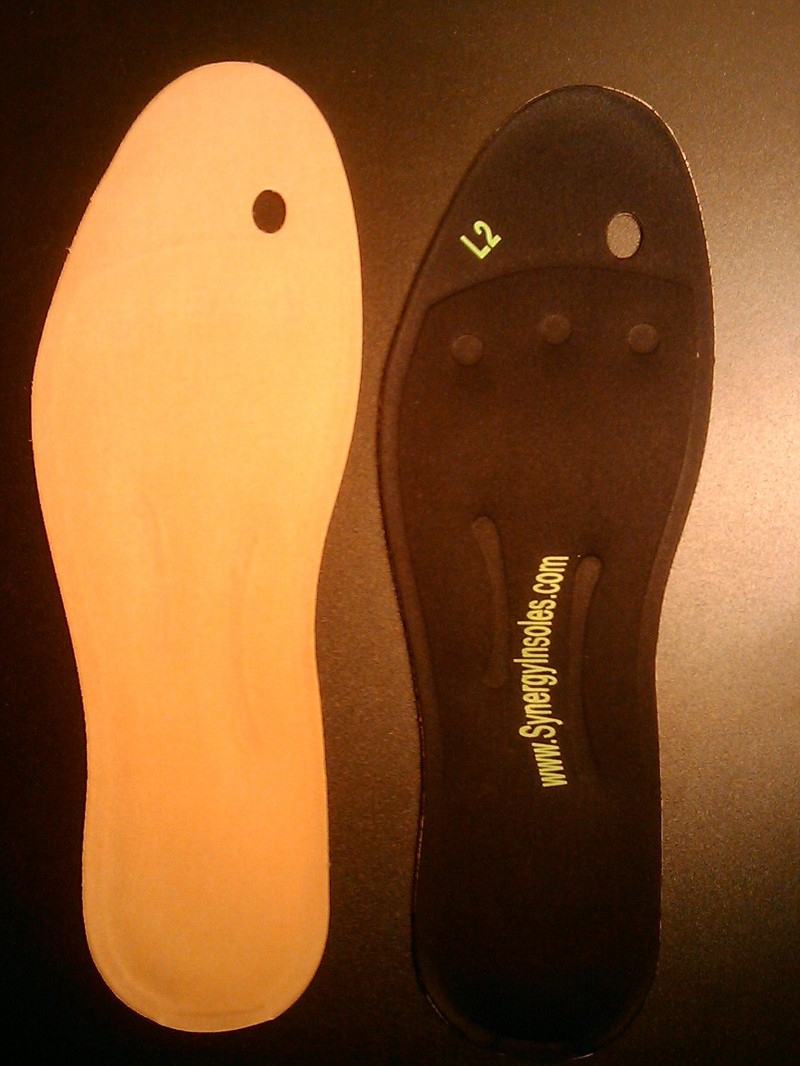
Recommended treatments, heel Spurs: cushioning for the heel is of little value. Your chiropodist/podiatrist may initially apply padding and strapping to alter the direction of stretch of the ligament. This is often successful at reducing the tenderness in the short term. Your chiropodist/podiatrist may suggest a course of deep heat therapy to stimulate the healing processes, allowing damage to respond and heal faster. In the long term, your chiropodist/podiatrist may prescribe special insoles (orthoses) to help the feet to function more effectively, thereby reducing strain on the ligaments and making any recurrence less likely. If pain from heel spurs continues, you may be referred to your GP who can prescribe an oral non-steroidal anti-inflammatory. Alternatively, localised hydrocortisone injection treatment may be given by your GP or an appropriate chiropodist/podiatrist. If pain persists, surgery may be considered. Heel Bursitis: in most cases, attention to the cause of any rubbing, and appropriate padding and strapping by your chiropodist/podiatrist will allow the inflammation to settle. If infection is present, your chiropodist/podiatrist will refer you to your GP for antibiotics. Heel Bumps: adjustments to footwear is often enough to make them comfortable. A leather heel counter and wearing boots may help. However, if pain persists, surgery may be necessary.

Surgical Treatment

Surgery to correct heel pain is generally only recommended if orthotic treatment has failed. There are some exceptions to this course of treatment and it is up to you and your doctor to determine the most appropriate course of treatment. Following surgical treatment to correct heel pain the patient will generally have to continue the use of orthotics. The surgery does not correct the cause of the heel pain. The surgery will eliminate the pain but the process that caused the pain will continue without the use of orthotics. If orthotics have been prescribed prior to surgery they generally do not have to be remade.

Prevention

Pain Under The Heel

Make sure you wear appropriate supportive shoes. Don’t over-train in sports. Make sure you warm up, cool down and undertake an exercise regime that helps maintain flexibility. Manage your weight, obesity is a factor in causing plantar fasciitis. Avoid walking and running on hard surfaces if you are prone to pain. You should follow the recognized management protocol “RICED” rest, ice, compression, elevation and diagnosis. Rest, keep off the injured ankle as much as possible. Ice, applied for 20 minutes at a time every hour as long as swelling persists. Compression, support the ankle and foot with a firmly (not tightly) wrapped elastic bandage. Elevation, keep foot above heart level to minimize bruising and swelling. Diagnosis. Consult a medical professional (such as a Podiatrist or doctor) especially if you are worried about the injury, or if the pain or swelling gets worse. If the pain or swelling has not gone down significantly within 48 hours, also seek treatment. An accurate diagnosis is essential for proper rehabilitation of moderate to severe injuries.

The Treatment And Cause Of Achilles Tendinitis

Overview

Achilles TendonitisAchilles tendinitis occurs when the band of tissue that connects the calf muscles at the back of the lower leg to the heel bone, the Achilles tendon, becomes inflamed. This condition is a result of overuse from intense exercise, jumping, running, and other activities that strain the tendon and calf muscles.

Causes

Achilles tendinitis can be caused by any activity that puts stress on your Achilles tendon. Tendinitis can develop if you run or jump more than usual or exercise on a hard surface. Tendinitis can be caused by shoes that do not fit or support your foot and ankle. Tight tendons and muscles, You may have tight hamstring and calf muscles in your upper and lower leg. Your tendons also become stiffer and easier to injure as you get older. Arthritis, Bony growths caused by arthritis can irritate the Achilles tendon, especially around your heel.

Symptoms

The Achilles tendon is a strong muscle and is not usually damaged by one specific injury. Tendinitis develops from repetitive stress, sudden increase or intensity of exercise activity, tight calf muscles, or a bone spur that rubs against the tendon. Common signs and symptoms of Achilles Tendinitis include, gradual onset of pain at the back of the ankle which may develop in several days up to several months to become bothersome. Heel pain during physical activities which may diminish after warming up in early stages, or become a constant problem if the problem becomes chronic. Stiffness at the back of the ankle in the morning. During inactivity, pain eases. Swelling or thickening of the Achilles tendon. Painful sensation if the Achilles tendon is palpated. If a pop is heard suddenly, then there is an increased chance that the Achilles tendon has been torn and immediate medical attention is needed.

Diagnosis

If Achilles tendonitis is suspected, avoid any exercise or activity that causes the pain. It is advisable to see a doctor promptly so that an accurate diagnosis can be made and appropriate treatment recommended. The doctor will take a full medical history and will ask about the nature and duration of the symptoms. They will perform a physical examination of the affected area. Ultrasound scanning may be used to assess damage to the tendon or surrounding structures. Occasionally MRI (magnetic resonance imaging) may be recommended. The symptoms of Achilles tendonitis are often similar to symptoms of other conditions such as partial Achilles tendon rupture and heel bursitis. This can make diagnosis difficult and a referral to an orthopaedic specialist may be required in order for an accurate diagnosis to be made.

Nonsurgical Treatment

Achilles tendonitis will often respond to rest or changes in activity, stretching, or ice after activity. Non-steroidal anti- inflammatory drugs (NSAIDs), such as ibuprofen or naproxen may also help. Physical therapy focusing on stretching and strengthening, massage, alternating hot and cold baths, and ultrasound or sound waves can also help with healing and comfort. The temporary use of a heel lift or the insertion of an arch support, called an orthotic, into the shoe or sneaker can also help. Although seldom necessary, the ankle may be kept in a short leg cast or splint. Surgery is rarely needed but can remove bone spurs or the bony prominence of the heel bone. The injection of corticosteroids such as cortisone into the area of the Achilles tendon is usually avoided because it may cause the tendon to rupture.

Achilles Tendon

Surgical Treatment

Surgery is considered when non-operative measures fail. Patient compliance and postoperative management is an important aspect of the operative management to prevent ankle stiffness or recurrence of the symptoms. Surgery usually requires a removal of the damaged tissue (debridement) and meticulous repair of the tendon. Post-operative immobilization is required, followed by gradual range of motion and strengthening exercises start. It may require 6 months for the full recovery. Some known complication are recurrence, stiffness of the ankle and deep vein thrombosis.

Prevention

You can take measures to reduce your risk of developing Achilles Tendinitis. This includes, Increasing your activity level gradually, choosing your shoes carefully, daily stretching and doing exercises to strengthen your calf muscles. As well, applying a small amount ZAX?s Original Heelspur Cream onto your Achilles tendon before and after exercise.